In the complex world of structural biology and drug discovery, understanding the precise three-dimensional structure of biological molecules and their interactions is paramount. High-resolution structural information provides critical insights into function, mechanism, and potential therapeutic targets. Two powerful techniques have revolutionized this field: Cryo-Electron Microscopy (Cryo-EM) and Micro-crystal Electron Diffraction (MicroED). ShuimuBio, an expert in structural biology services, offers advanced solutions leveraging these technologies to accelerate research and development.
Understanding MicroED: A High-Resolution Approach
Micro-crystal Electron Diffraction, or MicroED, is a cutting-edge technique specifically designed to determine high-resolution structures from samples that form tiny crystals – microcrystals or nanocrystals. Unlike traditional X-ray crystallography which requires larger, well-ordered crystals, MicroED can work with crystals that are often too small for X-ray diffraction. This capability is particularly valuable for researchers struggling to obtain suitable crystals for conventional methods.
MicroED operates by directing a beam of electrons at a frozen microcrystal or nanocrystal sample. As the electron beam passes through the crystal, it is diffracted, creating a pattern that contains information about the arrangement of atoms within the crystal. By collecting diffraction patterns from the crystal as it is tilted in the electron beam, scientists can computationally reconstruct a high-resolution three-dimensional model of the molecule.
This technique is especially well-suited for analyzing the structures of organic compounds. However, its applications extend beyond small organic molecules to include peptides and even proteins when they can be obtained in microcrystalline form. The ability of MicroED to work with minimal sample requirements and tiny crystals makes it an invaluable tool in structural determination.
The Connection: Cryo-EM and MicroED
While Cryo-EM Single Particle Analysis (SPA) is primarily used to resolve the structures of larger biological macromolecules like proteins and viruses that may not form ordered crystals, MicroED focuses on samples in a crystalline state, albeit microcrystalline. Both techniques involve preparing samples under cryogenic conditions to preserve their native state and using an electron microscope for imaging or diffraction.
A significant development highlighted in the sources is the integration of MicroED technology with conventional Cryo-EM systems. ShuimuBio has pioneered this integration with their proprietary eTasED software. This innovative software allows researchers to seamlessly apply MicroED techniques using standard Cryo-EM equipment without requiring any additional hardware modifications. This integration boosts the efficiency and accuracy of structural determination workflows, making high-resolution analysis more accessible.
By utilizing Cryo-EM systems for MicroED experiments, researchers can leverage the advanced electron optics and detection systems available on modern Cryo-EM platforms to collect high-quality diffraction data from microcrystals. This approach, which can be referred to as Cryo-EM MicroED in the context of performing MicroED on a Cryo-EM instrument, expands the capabilities of existing Cryo-EM facilities and offers a versatile platform for analyzing a wide range of sample types, from large, non-crystalline complexes via SPA to small molecules and peptides via MicroED.
Applications Across Research and Development
The power of MicroED, particularly when performed on integrated Cryo-EM MicroED systems, has far-reaching applications in various scientific disciplines, especially in the pharmaceutical industry and basic biological research.
· Small Molecule Drug Discovery: A primary application of MicroED is in the structure determination of small molecule drugs. Understanding the precise atomic arrangement of a small molecule is crucial for its design, synthesis, characterization, and understanding its interactions with biological targets. MicroED's ability to work with minimal amounts of microcrystalline material makes it highly suitable for samples obtained during early-stage synthesis or purification. It can provide definitive structural proof and insight into polymorphs or co-crystals. ShuimuBio applies MicroED for precise structural insights into small molecule samples.
· Peptide Structure Analysis: Peptides play vital roles in biological processes and are increasingly explored as therapeutic agents. Determining their three-dimensional structure is key to understanding their function and improving their properties. MicroED is capable of providing high-resolution structures of peptides, even from challenging samples. ShuimuBio uses MicroED for peptide crystal analysis.
· Protein Structure Resolution: While Cryo-EM SPA is the go-to method for many proteins, MicroED is valuable for proteins that form microcrystals. This is particularly useful for challenging proteins that don't grow into large, ordered crystals suitable for X-ray crystallography. Resolving protein structures at high resolution via MicroED provides detailed information about amino acid side chains, interactions, and binding sites, critical for understanding function and guiding drug design. ShuimuBio successfully delivers high-resolution protein structures using MicroED.
Illustrative Case Studies from ShuimuBio
ShuimuBio has demonstrated success in applying MicroED to various samples, achieving impressive resolutions:
· Proteinase K: Structure resolved at 1.50 Angstroms resolution for this 29.05 kDa protein. This showcases the ability of MicroED to resolve protein structures to near-atomic detail.
· FUS LC RAC1: Structure determined at 0.65 Angstroms resolution, 0.66 kDa peptide. Achieving sub-Angstrom resolution provides extremely precise details about the molecular structure.
· Acetaminophen: Structure resolved at 0.65 Angstroms resolution. While Acetaminophen is widely known as a small molecule drug, the source specifically describes the sample analyzed by MicroED as a peptide with a size of 0.66 kDa and a resolution of 0.65 Angstroms. Regardless of the classification provided in the source, achieving 0.65 Angstrom resolution on any sample demonstrates the exceptional resolving power of the MicroED technique as implemented by ShuimuBio.
These examples underscore ShuimuBio's proficiency in utilizing MicroED to obtain high-resolution structural data from different types of molecules, including proteins and samples described as peptides and small molecules.
Why Choose ShuimuBio for Your MicroED Needs?
ShuimuBio stands out as a leader in providing MicroED services, backed by significant expertise and technological advancements.
· Expert Team: ShuimuBio's core team includes excellent life and computing scientists and experts from the IT and pharmaceutical industries. Their MicroED team consists of PhD scientists from top institutions who are experts in both Cryo-EM and MicroED technologies. This deep expertise ensures that projects are handled with the highest scientific rigor.
· Proprietary Technology: The development of the eTasED software is a key advantage. This allows for the seamless integration of MicroED on conventional Cryo-EM systems, increasing efficiency and making the technology more accessible.
· Proven Success and High Resolution: ShuimuBio boasts a high success rate, having successfully delivered structures for over 80% of their MicroED projects. They have achieved resolutions ranging from 0.6 to 1.0 Angstroms, which is exceptional and provides atomic-level detail. This pursuit of extreme resolution is a stated advantage.
· Comprehensive "One-Stop" Services: ShuimuBio offers a holistic approach to structural biology projects. For MicroED, this includes initial project assessment, sample preparation guidance, data collection, and structure analysis. They even offer free project assessments to evaluate feasibility and potential risks. Beyond MicroED, their capabilities include Cryo-EM SPA, protein preparation and analysis, negative staining, cryo-characterization, and X-ray crystallography, providing a true "one-stop" solution from gene sequence to high-precision 3D structure. This integrated platform minimizes sample loss or degradation during transport between steps.
· Quality Control: ShuimuBio integrates strict quality control based on electron microscopy analysis and characterization throughout their protein preparation workflow to ensure samples are suitable for downstream structural analysis, including MicroED.
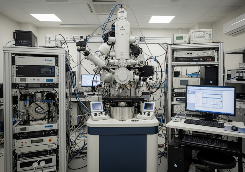
· Advanced Facilities: While MicroED can be performed on Cryo-EM systems, ShuimuBio's larger Cryo-EM platform and infrastructure support high-throughput and high-quality data collection. They have 8 high-end 300kV Cryo-EM instruments across Beijing and Hangzhou centers. Their facilities are maintained daily to ensure optimal performance.
Getting Started: Sample Submission for MicroED
To ensure the best results for MicroED analysis at ShuimuBio, specific sample requirements must be met:
· Sample Type: Small molecules, peptides, or proteins are accepted.
· Sample State: The sample must be in a stable crystalline form. This includes powder, lump, or other crystal states.
· Quantity: A minimum of 5mg of material is generally required. However, if 5mg is not feasible, a quantity that is visible to the naked eye may be sufficient.
· Stability: It is critical that the sample consists of stable crystals.
Researchers are advised to communicate with ShuimuBio's scientists or sales team at least 3 working days in advance for shipping arrangements.
Beyond MicroED: ShuimuBio's Broader Capabilities
While MicroED is a powerful technique for specific sample types, structural biology often requires a combination of methods. ShuimuBio's comprehensive service portfolio ensures that researchers can access the most appropriate techniques for their specific project needs.
Their Cryo-EM SPA services are crucial for resolving structures of larger molecules like antigen-antibody complexes, membrane proteins (GPCRs, ion channels, transporters), VLP, and multi-protein complexes that are difficult to crystallize. They have extensive experience in these areas.
Their protein preparation platform is a critical enabler, offering various expression systems (E. coli, mammalian, insect, cell-free) and purification methods (affinity, ion exchange, gel filtration, RP-HPLC) to obtain high-purity, homogeneous samples necessary for both Cryo-EM and MicroED. They specialize in difficult-to-express proteins, including membrane proteins.
They also offer analytical services like SPR, BLI, and ELISA to characterize protein-protein or protein-molecule interactions and perform quality control using SDS-PAGE, Western blot, and mass spectrometry. Negative staining and cryo-characterization services provide valuable preliminary data on sample quality and morphology before pursuing high-resolution methods.
This breadth of services, combined with their expertise in MicroED and Cryo-EM MicroED, positions ShuimuBio as a leading partner for comprehensive structural biology research.
Conclusion
Micro-crystal Electron Diffraction (MicroED) is a transformative technique for obtaining high-resolution structural insights from microcrystalline samples, particularly valuable for small molecules, peptides, and certain proteins. By enabling MicroED on standard Cryo-EM systems via innovative software like eTasED, ShuimuBio provides a powerful, efficient approach for structural determination.
Whether you are working on novel small molecule drugs, therapeutic peptides, or challenging protein targets, Cryo-EM MicroED offers a pathway to unlocking atomic-level structural details. With their expert team, advanced technology, high success rate, and integrated service platform, ShuimuBio is equipped to handle your most challenging structural biology projects.
To learn more about how MicroED, Cryo-EM MicroED, or other structural biology services can benefit your research, and to discuss your specific project needs, we invite you to visit https://shuimubio.com/.
Discover the power of high-resolution structure determination and accelerate your path to discovery with ShuimuBio.