Understanding the intricate three-dimensional structures of biological molecules is fundamental to advancing life sciences and drug discovery. Traditionally, obtaining high-resolution structural information for many complex or flexible biomolecules has been challenging. This is where cryo EM technology, specifically using the cryo electron microscopy principle, has emerged as a revolutionary tool, offering unprecedented insights into the molecular world.
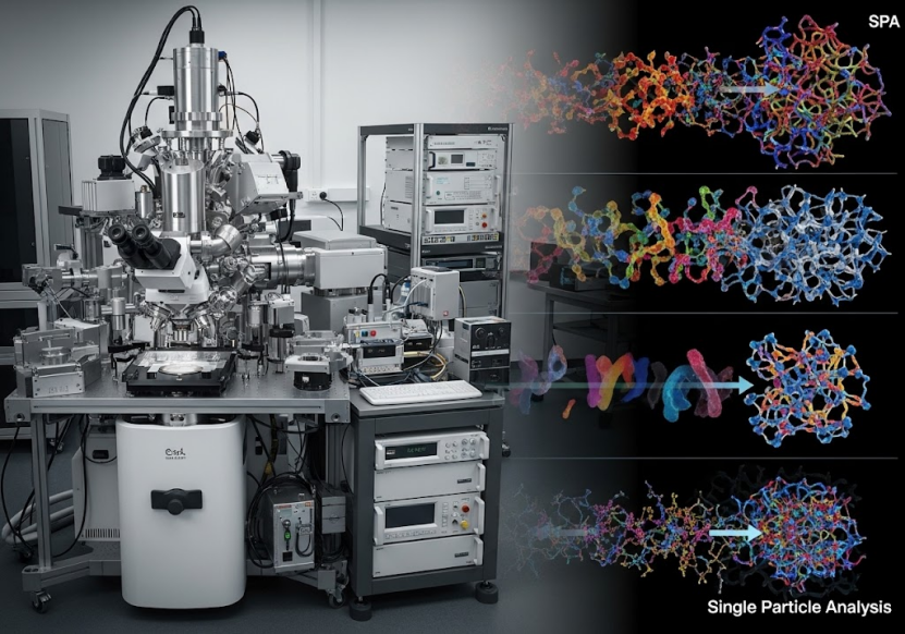
One of the most powerful applications within cryo EM technology is Single Particle Analysis (SPA). SPA is a technique that allows scientists to determine the high-resolution 3D structure of biological macromolecules such as proteins, viruses, and protein complexes, even if they are difficult or impossible to crystallize.
For researchers and companies seeking to leverage this advanced technology, understanding the core cryo electron microscopy principle behind SPA and the process involved is crucial. Let's delve into how SPA works.
The Core Principles of Cryo-EM Single Particle Analysis (SPA)
Single Particle Analysis (SPA) is a specialized method within cryo EM technology designed to reconstruct the 3D structure of biological samples by analyzing thousands or millions of individual images of identical particles. Unlike traditional electron microscopy, Cryo-EM requires samples to be preserved in a near-native, hydrated state by rapid freezing (vitrification). This avoids the damage and distortions that can occur with traditional staining or drying methods, which is a key cryo electron microscopy principle for preserving sample integrity.
The fundamental process of SPA involves several key steps:
1. Sample Preparation: This is a critical initial step. The biological sample (e.g., protein, virus, complex) must be purified and as homogenous as possible. Challenges like low sample concentration, strong background noise, preferred orientation, and damage at the air-liquid interface during freezing can hinder structure resolution. Specialized tools and methods, such as advanced protein purification techniques and innovative grid supports like GraFuture™ graphene-based grids, are employed to improve sample quality and overcome these issues. For SPA, specific sample requirements regarding concentration, volume, and purity must be met.
2. Cryo-Sample Freezing (Vitrification): A small volume of the purified sample is applied to a grid (often supported by a porous film) and rapidly plunge-frozen into liquid ethane or a similar cryogen. This process, called vitrification, freezes the water so quickly that ice crystals do not form, preserving the biological molecules in a thin layer of amorphous ice in their native state.
3. Cryo-Electron Microscopy Data Collection: The vitrified grid is transferred to a Cryo-EM microscope and kept at cryogenic temperatures (typically below -150°C). An electron beam is passed through the sample, and images are captured by a sensitive detector. Because biological samples are sensitive to electron radiation, low-dose imaging is used to minimize damage. Thousands or even millions of 2D projection images of individual particles, each oriented randomly on the grid, are collected. High-end cryo EM technology, like 300kV microscopes equipped with advanced detectors and energy filters, is essential for collecting high-quality data. Platforms offering 24-hour machine time service with advanced facilities ensure efficient data acquisition.
4. Image Processing and 3D Reconstruction: This is the computational heart of SPA. The millions of 2D images are processed using sophisticated algorithms.
o Particle Picking: Individual particle images are identified and extracted from the raw micrographs. AI-driven platforms can significantly automate and improve the efficiency of this step.
o 2D Classification: The extracted particle images are grouped into classes based on their similar orientations and appearances. This step helps to filter out damaged or aggregated particles and improve the signal-to-noise ratio.
o 3D Classification and Reconstruction: Images belonging to the same 2D classes are then used to reconstruct an initial low-resolution 3D map. This map is then refined using all selected particles, taking into account their determined orientations in 3D space. AI algorithms are increasingly used to enhance the efficiency and accuracy of data analysis and reconstruction.
o Refinement: The 3D reconstruction is iteratively refined to achieve the highest possible resolution.
5. Model Building and Validation: Finally, an atomic model of the biological molecule is built into the high-resolution 3D density map. The model is then refined and validated against the map and known biochemical data.
This multi-step process, leveraging the cryo electron microscopy principle of imaging vitrified samples and the computational power of image processing, allows for the resolution of structures ranging from moderately sized proteins (down to 51kDa) to large, complex assemblies like viruses and cellular machinery.
Why Use Cryo-EM SPA? Applications and Advantages
The advantages of SPA using cryo EM technology are numerous, making it invaluable in various research and development fields:
· Near-Native State Preservation: Samples are visualized in a hydrated, close-to-physiological state, offering a more accurate representation of their structure and function.
· Suitable for Challenging Samples: SPA is particularly effective for molecules that are difficult to crystallize, such as membrane proteins (GPCRs, ion channels, transporters), large complexes, and flexible proteins.
· Requires Less Sample: Compared to techniques like X-ray crystallography, SPA typically requires significantly less sample material.
· Captures Conformational Variability: By classifying images based on different appearances, SPA can reveal multiple functional conformations of a molecule or complex.
· Resolution: SPA can achieve near-atomic resolution, providing detailed insights into molecular interactions.
These advantages have led to widespread applications of cryo EM technology and SPA across various sectors:
· Drug Discovery:
o Target Identification and Validation: Resolving the structure of drug targets, including complex membrane proteins, is crucial for understanding their function and designing molecules that can interact with them.
o Mechanism of Action Studies: SPA helps researchers understand how drugs, including small molecules, peptides, and antibodies, bind to their targets and exert their effects.
o Antibody Drug Development: It is essential for resolving antibody-antigen complex structures, aiding in antibody design, optimization, and understanding neutralization mechanisms.
o Small Molecule Drug Development: Provides structural basis for designing highly selective and potent small molecules, supports fragment-based drug discovery (FBDD), and helps analyze biased ligands.
o PROTAC Research: Used to study the structural aspects of PROTACs binding to target proteins and E3 ligases.
· Vaccine Development: Cryo EM technology is used to resolve viral structures at high resolution, understand invasion mechanisms, study antibody-vaccine antigen interactions, and quickly analyze structures of new viral variants to inform vaccine design. It also plays a role in vaccine quality control by assessing particle morphology, size, integrity, and aggregation.
· Basic Research: Unraveling the structures of complex biological machinery (like ribosomes or spliceosomes), DNA/RNA structures and complexes, and various protein structures provides fundamental insights into biological processes.
· Nanomaterial Characterization: Used for characterizing the structure and morphology of nanoparticles, liposomes, LNPs, and AAV vectors.
ShuimuBio: A Leader in Cryo-EM SPA Services
As Asia's first commercial platform offering cryo EM technology services, ShuimuBio stands at the forefront of this field. Founded in 2017 with a core team of experts in structural biology, protein science, and computational biology, ShuimuBio provides comprehensive solutions.
ShuimuBio operates one of the world's largest commercial Cryo-EM platforms, featuring eight 300KV Cryo-EM microscopes strategically located in Beijing and Hangzhou. They have extensive experience, having completed over 400 Cryo-EM projects and resolved the structures of more than 150 proteins, achieving resolutions as high as 1.4 Å.
Their expertise covers the entire process from gene sequence to high-resolution 3D structure, offering a truly "one-stop" solution. This includes in-house protein expression and purification services capable of handling challenging targets like membrane proteins, negative staining and cryo-characterization for initial sample assessment, and high-quality data collection and advanced data analysis.
ShuimuBio leverages independent research and development in AI algorithms and software, such as the SMART software suite and the NanoSMART system for nanoparticle analysis, to boost efficiency and accuracy in image processing and data analysis. Their commitment to stringent quality control, often based on Cryo-EM analysis itself, ensures the delivery of high-quality results.
Beyond SPA, ShuimuBio also offers MicroED for micro/nano crystal structure resolution, machine time services, and a range of protein services including expression systems, purification, characterization, protein assays (SPR, BLI, ELISA), and a list of shelf proteins.
The successful application of ShuimuBio's platform and team has been documented in numerous high-impact publications, demonstrating their ability to resolve structures of various complex biological samples.
Get Started with Cryo-EM SPA
Engaging with cryo EM technology through a trusted service provider like ShuimuBio simplifies the complex process of structure determination. Their one-stop solution includes free project evaluation, flexible collaboration models, and regular project updates. By providing purified samples meeting specific requirements, researchers can access world-class facilities and expertise to accelerate their projects.
To learn more about how cryo EM technology and Single Particle Analysis can benefit your research or development project, and to explore the comprehensive services offered by ShuimuBio, please visit https://shuimubio.com/. Their team is ready to provide the structural insights you need to push the boundaries of science and drug discovery.