Understanding the intricate three-dimensional structure of proteins and other biological macromolecules is fundamental to unlocking their functions and developing targeted therapies. The field of structural biology is dedicated to this pursuit, employing various sophisticated techniques to visualize these tiny molecular machines. Among the most powerful and rapidly advancing protein structure determination methods, Cryo-Electron Microscopy (Cryo-EM), particularly Single Particle Analysis (SPA), has revolutionized the field.
Cryo-EM structural biology allows researchers to determine structures at near-atomic resolution, often overcoming limitations faced by traditional methods. For those seeking expert services in this critical area, exploring capabilities offered by leading platforms is essential. Visit https://shuimubio.com/ for more information on comprehensive structural biology services.
The Importance of Protein Structure
Proteins are the workhorses of the cell, involved in virtually every biological process. Their specific functions are directly dictated by their precise 3D shapes. Knowing a protein's structure can provide crucial insights into its mechanism of action, how it interacts with other molecules, and how diseases develop. This knowledge is indispensable for rational drug design, vaccine development, and fundamental biological research.
Diverse Protein Structure Determination Methods
The landscape of protein structure determination includes several key techniques, each with its strengths and applications.
· Single Particle Analysis (SPA) Cryo-EM: SPA is a powerful protein structure determination method that uses Cryo-EM technology to resolve the high-resolution 3D structure of biological macromolecules such as proteins and viruses. The process involves capturing numerous 2D images of purified particles frozen in a native-like state. Computer algorithms are then used to process and reconstruct these images into a high-resolution 3D structural model.
o Advantages of Cryo-EM SPA: Cryo-EM SPA offers several significant advantages. It allows samples to be kept in a state close to their native conformation. It can capture multiple conformational states, which is crucial for understanding dynamic proteins. This method often requires only small amounts of sample. Furthermore, it is effective in determining the structures of heterogeneous protein complexes. SPA is widely applied to study molecules like Protac, membrane proteins (GPCRs, ion channels, transporters), VLPs, peptides, and interactions between small molecules and target proteins.
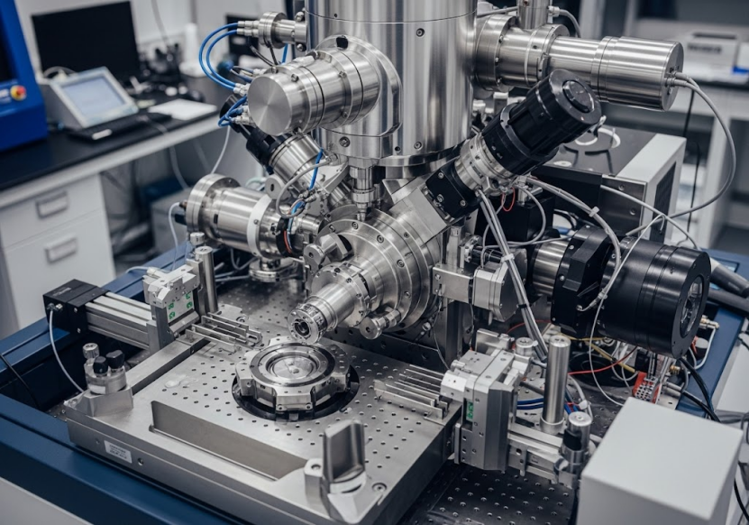
o Cryo-EM Facilities: High-quality Cryo-EM work requires state-of-the-art facilities. Leading platforms are equipped with top-tier microscopes and advanced computing platforms designed for high-quality structural resolution. For instance, some platforms boast a large network of 300kV Cryo-EM microscopes. These facilities often provide 24-hour data collection services, ensuring efficient project timelines. Regular maintenance is crucial to keep these instruments in optimal working condition, aiming for high availability and low fault rates.
o Challenges and Solutions: Traditional Cryo-EM sample preparation can face challenges such as air-liquid interface adsorption, severe preferred orientation, high sample concentration thresholds (>1μM), significant background noise, and difficulty in reconstructing structures of "small" macromolecules. To overcome these bottlenecks, innovative solutions like graphene support films (e.g., GraFuture™, GO, RGO) have been developed. These films can potentially solve preferred orientation issues and are suitable for samples with low molecular weight, low concentration, strong background noise, or where the air-liquid interface causes damage or preferred orientation.
o AI-Driven Platforms: Artificial intelligence (AI) is increasingly integrated into Cryo-EM workflows. AI-driven platforms utilize software (like SMART software series) to enhance data analysis efficiency, reduce machine runtime, and decrease the required data quantity. AI can also be used in cryo-characterization for automatically identifying nanoparticle features from images.
· MicroED (Micro-Electron Diffraction): MicroED is another cutting-edge protein structure determination method, specifically powerful for resolving high-resolution structures from microcrystals and nanocrystals, particularly for organic compounds, peptides, and proteins. This technique provides precise structural insights even from very small crystalline samples.
o MicroED Capabilities: Platforms with MicroED expertise can successfully deliver high-resolution structures, sometimes reaching resolutions of 0.6~1.0Å. The technology can be seamlessly applied using conventional Cryo-EM systems with original software (e.g., eTasED), requiring no additional system modifications, thus significantly boosting efficiency and accuracy.
· X-ray Crystallography: While Cryo-EM has gained prominence, X-ray crystallography remains a vital protein structure determination method. It involves growing crystals of the protein, then diffracting X-rays off the crystal lattice to generate a diffraction pattern that can be computationally converted into a 3D structure.
o Applications: X-ray crystallography can reveal high-resolution structures of molecules like antigen-antibody complexes, small molecule drugs, and peptides. It helps in understanding the dynamics of antigen-antibody interactions, optimizing antibody design, and improving the efficacy and specificity of antibody drugs. Platforms often offer "one-stop" services for crystal structure determination, covering everything from protein expression and purification to crystallization, data collection, and final structure resolution. Examples include resolving structures like KRAS and SARS-CoV M protein.
· Supporting Techniques: Complementary techniques are crucial in the structural biology workflow, particularly for sample quality control and initial characterization.
o Negative Staining & Negative Staining 2D: Negative staining is an electron microscopy technique that stains the background around the sample, making the biological structure visible. It is useful for quickly obtaining low-resolution 2D projection images of macromolecules and complexes, providing preliminary information on particle size, uniformity, oligomeric state, morphology, particle density, protein structure, flexibility, sample integrity, and conformational heterogeneity. Negative staining 2D specifically refers to analyzing these samples using 2D imaging, often for structures arranged in a 2D plane. These methods are commonly used for observing viruses, nanoparticles, organelles, and protein complexes.
o Cryo-Characterization: Cryo-characterization utilizes ultra-low temperature technology to maintain samples in their natural state for high-resolution observation and analysis. It is particularly advantageous for observing the structures of proteins, liposomes, exosomes, and material interfaces. AI systems can assist in automatically identifying nanoparticle features like size distribution, circularity, lamellar structure (full/empty), and integrity from Cryo-EM images. This is applied in research involving LNPs, liposomes, AAVs, and other viral vectors.
o Protein Quality Control and Analysis: Ensuring high sample purity and homogeneity is paramount for successful structure determination. Techniques used for protein quality control include SDS-PAGE, Western blot, mass spectrometry analysis, thermal stability, and solubility tests. Protein-protein or protein-molecule binding analysis can be performed using methods like SPR, BLI, MST, and ITC. SPR (Surface Plasmon Resonance) and BLI (Bio-Layer Interferometry) are widely used for detecting binding kinetics parameters between proteins and other molecules, essential for drug discovery and protein engineering. ELISA (Enzyme-Linked Immunosorbent Assay) is used for quantitative analysis of target protein content based on antigen-antibody reactions. These techniques provide crucial data to support structural studies.
The Importance of Sample Preparation
Regardless of the structural determination method used, the quality of the biological sample is critical. High-quality protein is the foundation for method development, compound screening, and structural biology. Expert platforms offer comprehensive protein preparation and analysis services, including various protein expression systems (E. coli, mammalian cells, insect cells, cell-free systems) and purification methods (affinity chromatography, ion exchange chromatography, gel filtration chromatography, RP-HPLC). These services aim to produce target proteins with high purity and activity, minimizing issues like aggregation or misfolding that can hinder structural studies. Overcoming the challenges of preparing difficult-to-express proteins is key to improving structural resolution capabilities. Strict quality control measures, often based on Cryo-EM analysis, are applied to ensure samples meet the requirements for downstream research.
Applications in Drug Discovery and Development
Cryo EM structural biology and other protein structure determination methods are indispensable in modern drug discovery and development across various therapeutic areas:
· Vaccine Development: Cryo-EM plays a crucial role in vaccine development by resolving the 3D structures of viruses at near-atomic resolution, aiding in understanding viral invasion mechanisms and informing vaccine design. Examples include structural studies of SARS-CoV-2 spike protein and its complexes with receptors, crucial for understanding infection and designing vaccines. Cryo-EM is also used for vaccine quality control, assessing particle morphology, size, integrity, and aggregation during manufacturing. It helps study the interaction mechanisms between antibodies and vaccine antigens to optimize immunogenicity. Furthermore, the ability to quickly resolve structures of new viral variants using Cryo-EM is vital for adapting vaccine strategies in response to outbreaks.
· Antibody Drug Development: Cryo-EM has significant value in antibody drug research. It enables the resolution of high-resolution 3D structures of antibody-antigen complexes, helping researchers understand recognition mechanisms and binding sites. This structural information is critical for designing more effective antibody drugs. Cryo-EM is also used to study antibody drug action mechanisms, including binding and signaling pathway modulation. It supports antibody optimization and design by revealing interaction dynamics and conformational changes. The ability to resolve structures of challenging targets like membrane proteins (e.g., GPCRs) is particularly valuable. Overall, Cryo-EM accelerates the antibody drug development process by providing detailed structural information efficiently.
· Small Molecule Drug Discovery: Cryo-EM is a vital tool in small molecule drug research. It helps resolve high-resolution structures of drug targets (like membrane proteins and enzymes), aiding in understanding where small molecule drugs act. By resolving complex structures of targets bound to small molecule ligands, researchers gain detailed insights for designing highly selective and effective drugs. Cryo-EM is also valuable in studying the interaction mechanisms between small molecule drugs and their targets. It shows great potential in fragment-based drug discovery (FBDD) by revealing detailed interactions between small molecule fragments and protein targets. Similar to antibody drug development, Cryo-EM's efficiency helps accelerate the overall drug discovery process. It is also uniquely advantageous for studying biased ligands that selectively modulate specific signaling pathways mediated by targets like GPCRs. The technique's ability to resolve complex target structures is highly beneficial.
Why Choose a Specialized Platform?
Engaging with a specialized platform for cryo em structural biology and protein structure determination methods offers distinct advantages. Such platforms consolidate extensive experience, cutting-edge technology, and expert teams. For example, a platform founded by leading life and computational scientists may have resolved over 150 protein structures with resolutions as high as 1.8 Angstroms and handled over 400 Cryo-EM projects. They often provide comprehensive, one-stop services, from sample preparation to data delivery, saving time and resources. Free project evaluation and regular progress meetings ensure client satisfaction and project success.
Leading platforms possess advanced Cryo-EM facilities specifically designed for high-quality structural resolution. They are staffed by expert scientists with doctoral degrees specializing in structural biology, protein science, and computational biology. Their rich experience spans diverse sample types, from membrane proteins to antigen-antibody complexes. A commitment to achieving the highest possible resolution is a hallmark of top providers. Innovative technologies like proprietary AI algorithms and specialized consumables (like graphene grids) can significantly boost structure resolution efficiency and precision.
Conclusion
Cryo EM structural biology, alongside other advanced protein structure determination methods, is continuously pushing the boundaries of our understanding of biological systems. These powerful techniques are fundamental to basic research and have profound implications for the development of new vaccines and therapeutic drugs. By providing detailed insights into the molecular architecture of life, they pave the way for scientific breakthroughs and medical advancements.
Platforms specializing in these services are equipped with the necessary expertise, technology, and infrastructure to tackle challenging projects and deliver high-resolution structural data. Whether the goal is to determine the structure of a novel drug target, understand the mechanism of a potential therapy, or ensure the quality of a biological product, leveraging these advanced methods is essential.
To learn more about comprehensive services in cryo em structural biology and diverse protein structure determination methods, and to see how these capabilities can support your research or development needs, we encourage you to visit https://shuimubio.com/.